2022.10.24 | Questions 1-2
Barth syndrome & heterotaxy
Questions
Question 1
A 2 year old boy presents with recurrent pneumonia, cardiomyopathy, and neutropenia. Genetic sequencing is performed and reveals a variant in TAFAZZIN. The synthesis of which type of lipid, present only in the inner mitochondrial membrane, is affected in this disorder?
Question 2
A woman at 32 weeks gestation presents with concern for fetal heterotaxy on prenatal ultrasound. An MRI is performed to further characterize fetal anatomy. Which of the following fetal MRI findings is most likely to be consistent with a diagnosis of heterotaxy?
Explanations
Question 1
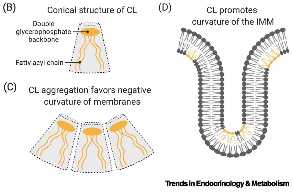
Cardiolipin is the correct answer. Cardiolipin (CL) comprises 20% of all lipid in the inner mitochondrial membrane (IMM) and is not found in other cellular membranes. The IMM contains cristae, which are tightly-curved IMM folds enriched in CL (panel D). The conical structure of CL (panel B-C) cause the membrane to curve. The TAFFAZIN protein adds a type of fatty acid called linoleic acid (C18:2) to cardiolipin, allowing the cardiolipin to mature and perform its function in maintaining the shape of the mitochondria.
Pathogenic variants in TAFFAZIN cause Barth syndrome, an X-linked disorder associated with cardioskeletal myopathy and neutropenia, the latter of which can predispose to recurrent infections. 💡Think “Barth affects Boys.” The prefix “cardio-” in the word “cardiolipin” reflects the fact that CL is present in high concentration in the heart; it should therefore make sense that Barth is associated with cardiomyopathy.
The lipids listed in the other 3 choices (cholesterol, phosphatidylethanolamine, phosphatidylcholine) are found in membranes outside of the mitochondria.
Learning objective
Cardiolipin is uniquely present in the inner mitochondrial membrane and requires tafazzin for proper synthesis. Mutations in TAFAZZIN result in Barth syndrome.
2023 ABMGG General Exam Blueprint | V. Single Gene Inheritance → d. Single Gene Disorders → ix. Metabolic disease → 7. Mitochondrial disorders (page 3)
Question 2
Patients with heterotaxy often have splenic abnormalities such as asplenia or polysplenia (multiple small spleens). The arrangement of the other organs described (leftward-pointing heart, left-sided gastric bubble, tri-lobed right lung) is consistent with normal human anatomy.
Heterotaxy is a term derived from two Greek words: hetero- (meaning “different”) and “-taxy” (meaning “arrangement”). The word “heterotaxy” describes situations in which the internal thoracic and abdominal organs are “arranged differently” along their left-right axis. The organs themselves may also lose their natural left-right asymmetry, resulting in isomerism.
Isomerism is defined as an unusual symmetry of normally asymmetric visceral organs (e.g. heart, liver). There are two types of isomerism: right atrial isomerism (RAI) and left atrial isomerism (LAI), referring to which atrial appendage is duplicated. In LAI, structures on the left side of the body are duplicated, resulting in major cardiac defects (e.g. having two left atria) and polysplenia. In contrast, patients with RAI often have asplenia, as right-sided structures are duplicated while the left-sided viscera may be absent.
The most clinically significant consequence of heterotaxy is abnormal cardiac development, which can lead to life-threatening structural cardiac disease or cardiac conduction anomalies (e.g. heart block). Bile duct anomalies associated with an abnormally symmetric liver can also be seen, leading to biliary atresia. A small fraction of patients have heterotaxy secondary to Primary Ciliary Dyskinesia (PCD), though a genetic basis for heterotaxy is not identified in most cases.
Learning objective
Heterotaxy is characterized by an unusual symmetry and arrangement of internal organs. Organs that are normally present on only 1 side of the body (eg spleen) may be duplicated or absent.
2023 ABMGG General Exam Blueprint | V. Single Gene Inheritance → d. Single Gene Disorders → x. Other (page 3) | VII. Reproductive Genetics (page 4)


